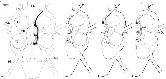
Fig. 1.
Anatomy of the acoustic afferents of the right bulba acustica in Ormiaochracea.A, This is a dorsal view of az-projection of 28, 1-μm-thick confocal optical sections. Texas Red dextran dye was applied to the bulba acustica, and >50 afferents were stained. Stained afferents project to three acoustic neuropil regions in the ventral region of the three neuromeres in the fused thoracic ganglion of the fly. B, A single auditory afferent stained with fluorescent dextrans. There is a dense projection into the T2 auditory neuropil area from this afferent. The T1 and T3 neuropil regions receive minimal projections from this afferent. C, A single auditory afferent stained with fluorescent Texas Red dextrans. There are dense projections into the T1 and T2 auditory neuropil area from this afferent. The T3 neuropil region does not receive any projections from this afferent.D, A single auditory afferent stained by Lucifer yellow injection. A single branch projects into the T1 auditory area from the primary neurite. This branch then continues projecting to the T2 auditory neuromere. There is a separate projection from the primary neurite into the T2 auditory area and a minimal projection to the T3 auditory area. AMN, Accessory mesothoracic neuromere;CN, cervical connective; FN, frontal nerve; HN, haltere nerve; T1, prothoracic neuromere; T2, mesothoracic neuromere;T3, metathoracic neuromere; VE, ventral ellipse; WN, wing nerve.

