
Fig. 10.
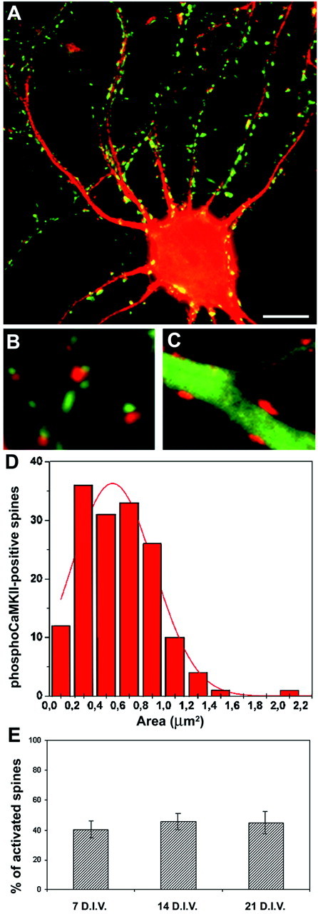
Postsynaptic CaMKII phosphorylation in stage V neurons. A, Stage V embryonic hippocampal neurons at 21 DIV were exposed to KCl (55 mm) for 1 min, fixed, and processed for double immunofluorescence with anti-synapsin (green) and phosphospecific anti-CaMKII (red) antibodies. B, C, High-magnification images of distal dendrites treated as inA and processed for double immunofluorescence with phosphospecific anti-CaMKII (red) and either anti-synapsin (B) or anti-MAP2 (C) antibodies (green). Note the absence of labeling for phosphoCaMKII in the dendritic shaft. The phosphoCaMKII-positive spots are always juxtaposed to synapsin-positive, presynaptic terminals and confined to MAP2-negative areas, defining them as spines. D, Size distribution of the phosphoCaMKII-positive areas in distal dendrites. The continuous red line represents the Gaussian fitting of the curve. E, Stage V embryonic hippocampal neurons at 7, 14, and 21 DIV were processed as in A. The histograms show the percentage of phosphoCaMKII-positive spines (mean ± SD) at each stage. The number of activated spines was normalized for the number of synapsin-positive presynaptic boutons present in each field. Scale bars: A, 6 μm; B, C, 2 μm.