Fig. 7.

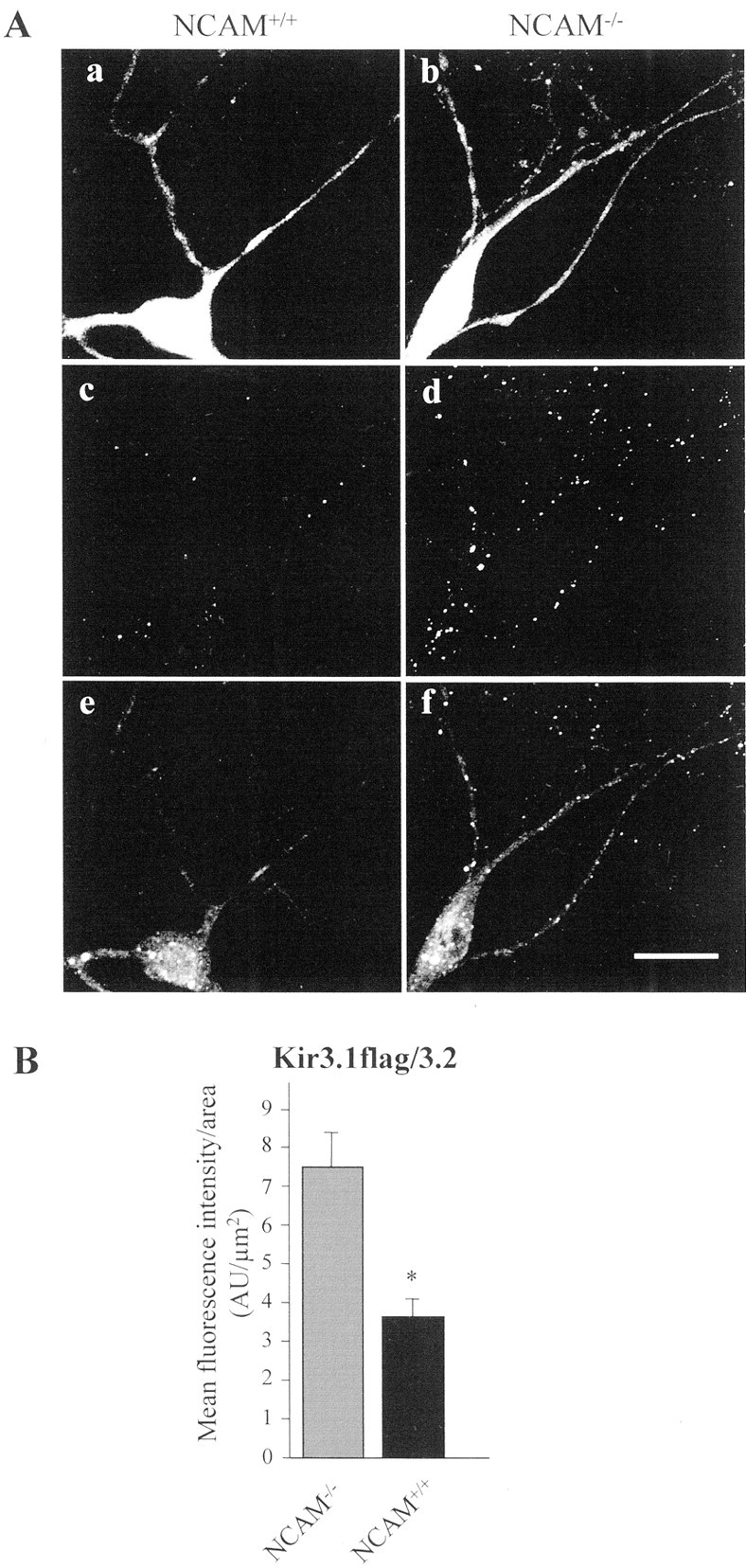
Kir3.1flag/3.2 channels are enriched at the cell surface and in the dendrites of NCAM−/− neurons.A, Confocal images of NCAM−/−(a, c, e) and NCAM+/+ (b, d, f) neurons cotransfected with Kir3.1flag/3.2 channels and GFP. Only cells expressing comparable amounts of GFP were selected for quantification (a, b). Neurons were stained with flag antibody before fixation (c, d). After permeabilization, cells were stained with Kir3.1 antibody to visualize intracellular Kir3.1flag/3.2 channels (e, f). In NCAM−/− neurons, more flag-immunoreactive clusters are detectable on the cell surface than in NCAM+/+neurons (d vs c), demonstrating that more Kir3.1flag/3.2 channels are present at the cell surface in NCAM−/− neurons. In NCAM+/+neurons, intracellular Kir3.1 immunoreactivity is prominent in the cell soma around the nucleus, whereas it is only weakly present in neurites (e). In contrast, NCAM−/−neurons show a more diffuse intracellular Kir3.1 immunoreactivity in neurites and cell soma (f).B, Bar graph showing in arbitrary units (AU) the mean fluorescence signal ± SEM of cell-surface flag immunostaining of Kir3.1flag/3.2-transfected NCAM−/− (7.45 ± 0.96 AU) and NCAM+/+ (3.7 ± 0.53 AU) neurons (n = 30 each), demonstrating that Kir3.1flag/3.2 surface localization is reduced by ∼50% in NCAM+/+ versus NCAM−/− neurons. Scale bar, 20 μm. ∗Statistically significant difference between genotypes (Student's t test; p < 0.01).
