Fig. 4.
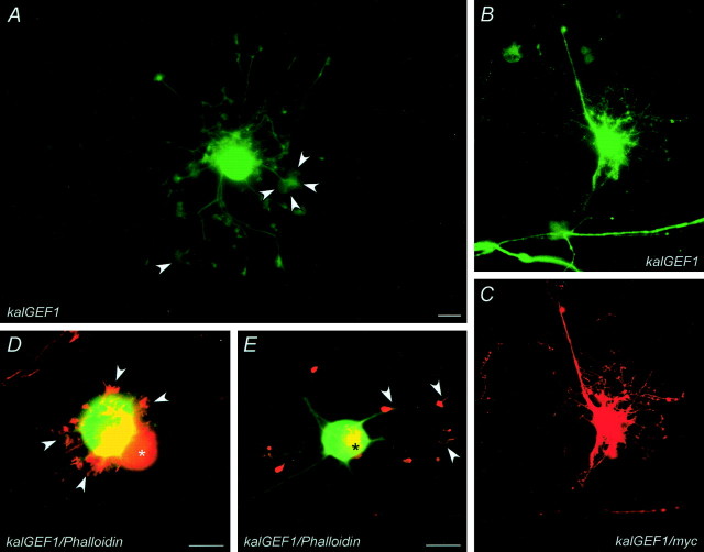
KalGEF1 domain expression is key to the Kalirin-induced fiber outgrowth phenotype. A, Microinjection of sympathetic neuron with 200 ng/μl kalGEF1 expression vector induced robust fiber outgrowth and branching (compare with Fig. 2A). Growth cones at distal fiber terminals appeared as broad lamellipodial sheets (arrowheads). B, C, A neuron injected with kalGEF1 and EGFP (B) and processed immunocytochemically to visualize the myc-epitope of the kalGEF1construct (C) demonstrated kalGEF1 expression in newly formed fibers and terminals. D, E, KalGEF1-induced fiber initiation resulted from extensive actin cytoskeleton reorganization. Micrographs of two different kalGEF1/EGFP-injected neurons (green) were merged with micrographs of the same neurons visualized with TRITC-phalloidin (red). The noninjected sympathetic neurons (D,asterisk) displayed relatively uniform staining for filamentous actin. Expression of kalGEF1 caused a redistribution of filamentous actin to emerging fiber outgrowths (D;arrowheads mark red lamellipodial filigree emerging from green microinjected neuron) and to large aggregates in the perinuclear region of the cell soma (yellow represents actin aggregates fromgreen and red fluorescence overlay). At later stages of fiber development the kalGEF1-injected neurons typically displayed prominent staining for filamentous actin (red) at growth cones (E, arrowheads); aggregates of filamentous actin were still apparent in neuronal soma (asterisk).

