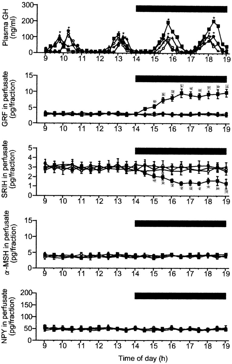
Fig. 1.

Representative profiles of plasma GH in four fed male rats and group data of neurohormones in the ME–ARC perfusates in the four fed groups before and during the leptin infusion. The number of animals in each group was 9–11. In this figure and Figure 3, (1) the time of the perfusate collection for neuropeptide assays is shifted 15 min ahead of the actual time of perfusion, because the dead space of the pull system (225 μl) corresponds to a 15 min period of perfusion (flow rate, 15 μl/min); (2) data of the four neuropeptides in perfusates are expressed as point values at the center of their collection periods; and (3) where SE values are not shown, they were smaller than the symbols. Black bar, Period during which leptin or aCSF (vehicle) was infused; filled squares, leptin (10 ng/ml); filled triangles, leptin (3.0 ng/ml);filled circles, leptin (1.0 ng/ml); open circles, aCSF (control); stars, significant GH pulses as detected by Cluster analysis; dotted crosses, statistically significant versus the other three groups.
