
Fig. 1.
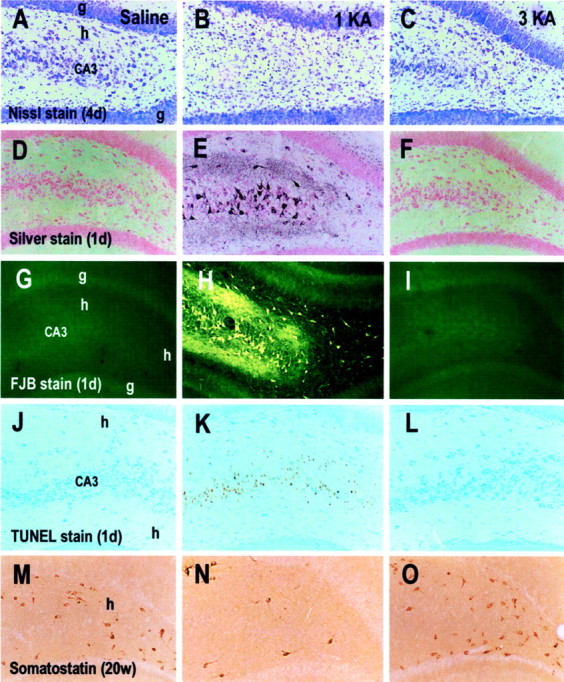
Microphotographs showing cresyl violet (Nissl stain) stain (A–C), silver stain (D–F), Fluoro-Jade B (FJB) stain (G–I), TUNEL stain (J–L), and somatostatin immunohistochemical stain (M–O) in rats treated with saline (A, D, G, J, M), a single KA (1KA) (B, E, H, K, N), and triple KA (3KA) (C, F, I, L, O) injections 4 d (4d) (A–C), 1 d (1d) (D–L), and 20 weeks (20w) (M–O) after the last saline or KA injection. Adjacent sections (D, E, G, J, H, K, orF, I, L) were obtained from the same brains.CA3, CA3 pyramidal cell layer; g, granule cell layer of the dentate gyrus; h, hilus. Magnification: 35× for all microphotographs.