
Fig. 4.
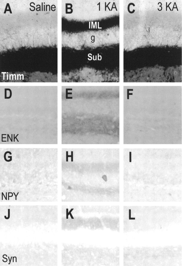
Microphotographs showing Timm stain (Timm) (A–C) and immunohistochemical stain for enkephalin (ENK) (D–F), neuropeptide Y (NPY) (G–I), and synaptophysin (Syn) (J–L) in rats receiving injections of saline (A, D, G, J), a single KA (1KA) (B, E, H, K), or triple KA (3KA) (C, F, I, L) 20 weeks after the last saline or KA injection. Adjacent sections (A, B, D, E, G, H, K, J, and C, F, I, L) were obtained from the same brains. g, Granule cell layer of the dentate gyrus; IML, inner molecular layer of the dentate gyrus; sub, subgranule layer of the dentate gyrus. Magnification: 70× for all microphotographs.