
Fig. 1.
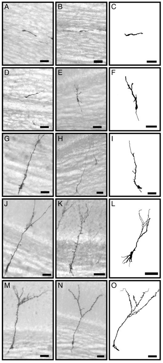
Alkaline phosphatase staining of virus-infected newly generated cells in the GCL of the olfactory bulb. Photomicrographs (left and center columns) and camera lucida drawings (right column) show examples of stained developing granule neurons at different maturation stages as defined in Results: class 1 (A–C); class 2 (D–F); class 3 (G–I); class 4 (J–L); class 5 (M–O). Scale bars, 25 μm.