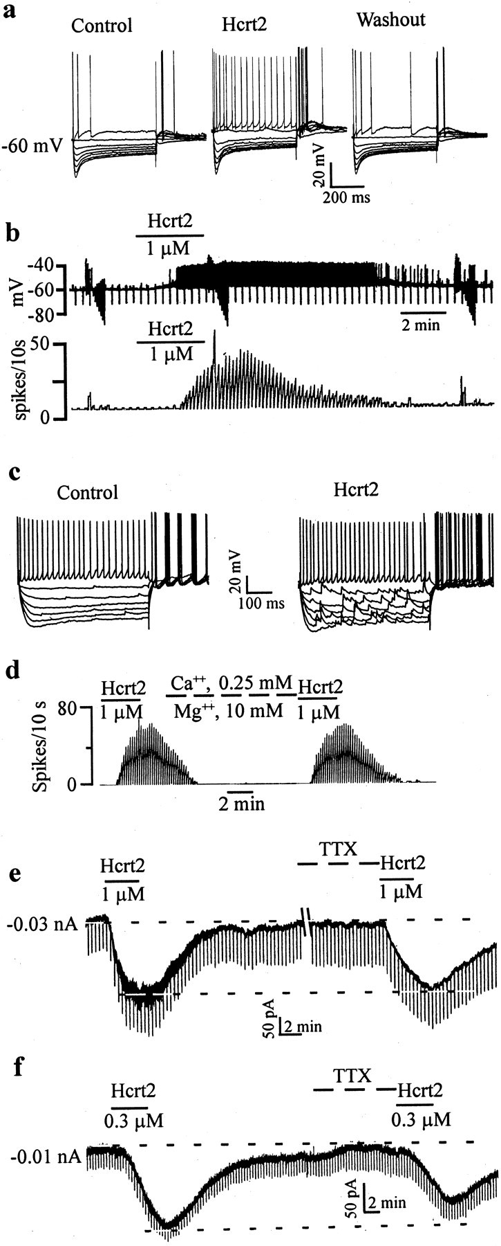
Fig. 2.

GABA-type MSDB neurons are excited by Hcrt2 via a direct postsynaptic effect. a, Intracellular recordings from a GABA-type MSDB neuron showing the reversible, excitatory effect of bath-applied Hcrt2. Note the depolarizing sag in response to the hyperpolarizing pulses and the presence of an anode-break excitation after termination of the hyperpolarizing pulses, features that are characteristic of GABA-type MSDB neurons. This cell had a spike duration of 0.43 msec and was also excited by muscarine (data not shown). b, Chart records showing the membrane potential (top) and firing rate (bottom) of a GABA-type MSDB neuron recorded intracellularly. Note that Hcrt2 depolarized the cell and induced firing [action potentials clipped, see rate record (bottom trace)]. c, A current-clamp recording from a GABA-type MSDB neuron that also showed a clear-cut increase in synaptic activity after bath application of Hcrt2. d, The effect of Hcrt2 in normal ACSF and after blockade of synaptic transmission using ACSF containing 0.5 mm Ca2+ and 10 mm Mg2+. e,f, Voltage-clamp recordings from two GABA-type neurons (holding potential, 65 mV; step command, −5 mV every 20 sec) show that Hcrt2 produced inward currents, 210 and 140 pA, respectively. Also note the increase in synaptic noise in e. Note that, although the Hcrt2 effect persisted even after bath application of 2 μm TTX in both neurons, the response to Hcrt was reduced in the cell shown in f (before TTX, 140 pA; after TTX, 80 pA) but not in the cell shown in e; five of nine neurons tested showed a reduction in the Hcrt response after application of TTX. Also note that TTX blocked the Hcrt-induced increase in synaptic noise. This may indicate the presence of a TTX-sensitive sodium component and/or an indirect component (see Discussion).
