
Fig. 1.
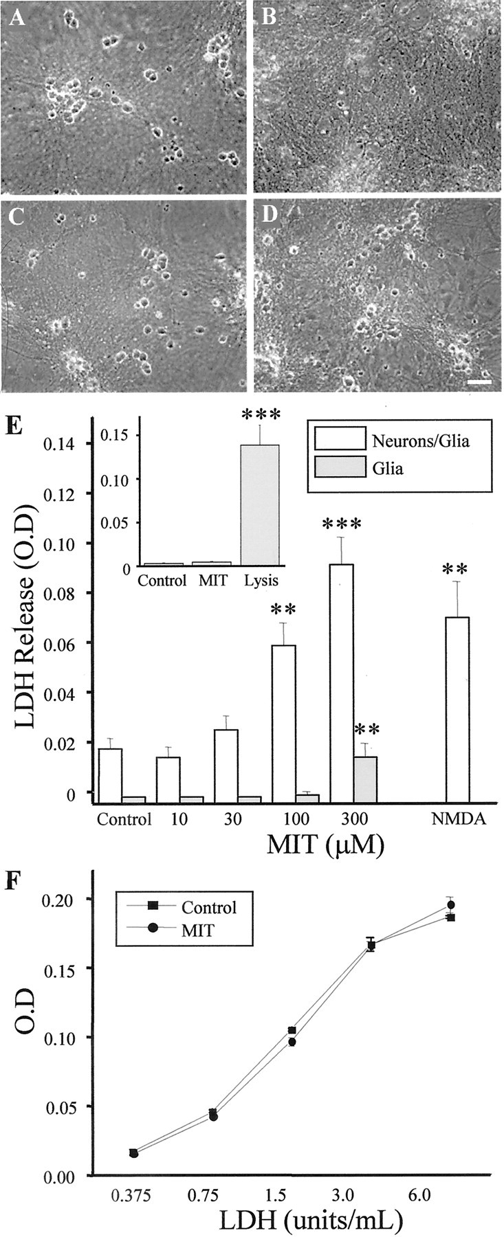
MIT is neurotoxic in vitro.A–D, Phase–contrast micrographs of rat cortical cultures 24 hr after being treated for 10 min with either vehicle (A), 100 μm MIT (B), 10 μm TPEN (C), or 100 μm MIT plus 10 μm TPEN (D). Note the relative absence of phase–bright (live) neurons in B and the neuroprotective actions of TPEN in D. E, Concentration–toxicity relationship for MIT in control mixed cultures (Neurons/Glia) and in sister cultures that had been treated 72 hr earlier with kainic acid (1 mm, 24 hr) to remove the neuronal component (Glia). MIT results in a large increase in LDH release (an index of cell death) in the mixed but not in the glial cultures. LDH release induced by a 1 hr exposure to 200 μm NMDA (a selective neuronal toxin) is included for comparison. Values represent the mean ± SD for a total of six experiments in the mixed cultures and three experiments in the kainate-treated cells; ∗∗p < 0.01; ∗∗∗p < 0.001. The inset shows a representative experiment, in triplicate (mean ± SD), performed on primary mouse astrocytes. A 10 min exposure to 100 μmMIT was not toxic to the cells. Total LDH in the culture was measured after cell lysis; ∗∗∗p < 0.001.F, LDH activity measured with known concentrations of the enzyme alone or in the continuous presence of 100 μmMIT. No differences were observed between the two standard curves. Values represent the mean ± SD of three independent measurements.