
Fig. 1.
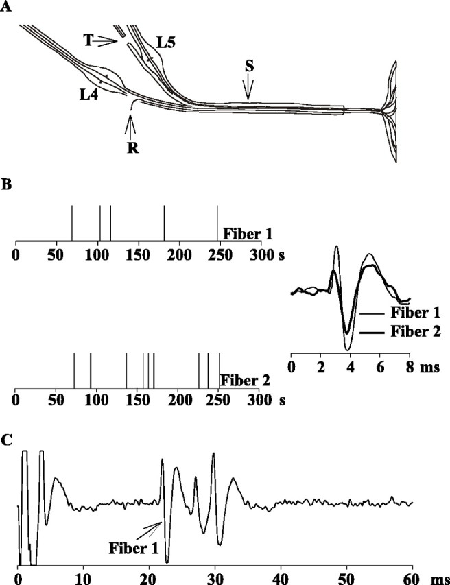
Spontaneous activity in two typical C-fiber afferents recorded 8 d after an L5 ventral rhizotomy.A, Teased-fiber techniques were used to record activity from single nerve fibers of the L4 spinal nerve (R) in sham-operated animals and in animals after transection of the L5 ventral root (T). Electrical stimulation of the sciatic nerve (S) was used to identify and count the number of C-fibers at the recording electrode. B, The presence of spontaneous action potential activity was assessed over a 5 min recording interval. In this example, two C-fibers with low-grade spontaneous activity were recorded simultaneously. On the left, eachpanel represents the spontaneous activity of one fiber, and each vertical line corresponds to the time of occurrence of an action potential. The action potential waveforms are illustrated on the right. C, Suprathreshold electrical stimulation at the sciatic nerve produced three discrete action potential waveforms at C-fiber latencies. The action potential waveform starting at 21 msec (peak at 22 msec) had the same shape as the spontaneously active fiber 1, providing evidence that the spontaneous activity came from this C-fiber. The two other C-fibers had superimposed waveforms (from 26 to 32 msec). Fiber 2 corresponds to one of these C-fibers.