
Fig. 1.
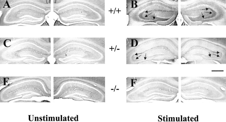
Reduction of seizure-induced increase of p-trk immunoreactivity in trkBshcmutant mice.A, C, E, p-trk immunoreactivity in unstimulated +/+, +/−, and −/− mice. Note the absence of detectable immunoreactivity in dentate hilus and CA3 stratum lucidum of hippocampus. B, D,F, p-trk immunoreactivity in +/+, +/−, and −/− mice 24 hr after a seizure evoked by stimulation of right amygdala.Arrows denote immunoreactivity in dentate hilus and stratum lucidum in stimulated +/+ and +/− mice. Despite some differences in background among individual animals, no systematic variation in background attributable to genotype or stimulation status was present (data not shown). Note that quantitation was controlled for individual differences in background by use of a control region (corpus callosum) intrinsic to each section. Scale bar, 650 μm.