
Fig. 1.
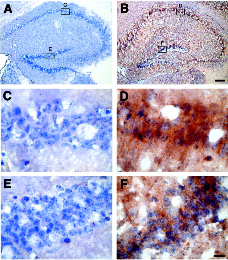
Immunohistochemistry for TTR in the hippocampus of control and APPSw overexpressing mice. Sections from nontransgenic mice (A, C, E) and APPSw mice (B, D, F) were immunostained for TTR and counterstained with hematoxylin. TTR is increased throughout the hippocampus in APPSw mice (B) compared with control mice, which contain little to no TTR (A). TTR levels in APPSw mice are largest around the neurons in CA1 (D) and the dentate gyrus (F). There is virtually no immunostaining for TTR in CA1 (C) and the dentate gyrus (E) of nontransgenic mice. Scale bars: (inB) A, B, 150 μm; (inF) C–F, 10 μm.