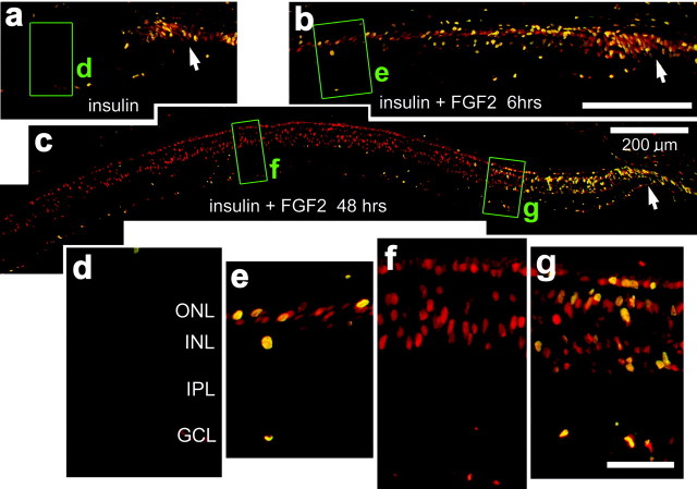
Fig. 1.
Exogenous insulin and FGF2 stimulate the proliferation of cells within peripheral regions of the retina. Vertical sections of the retina were labeled with antibodies to PCNA (in red) and BrdU (in green). Cells double labeled for PCNA and BrdU appear yellow, because all BrdU-labeled cells express PCNA. a–c, Montage images of retinas from eyes that received three consecutive daily injections of BrdU with insulin (a, d) or insulin with FGF2 (b, c,e–g). Injections were made from P7 through P9, and eyes were harvested 6 hr (b, e), 24 hr (a, d), or 48 hr (c,f, g) after the final injection.d–g, Enlarged images of the areas boxed out ingreen in a–c. Arrows, Retinal margin. Scale bars: b (for a,b), c (for d–f), 200 μm; g, 50 μm. GCL, Ganglion cell layer.

