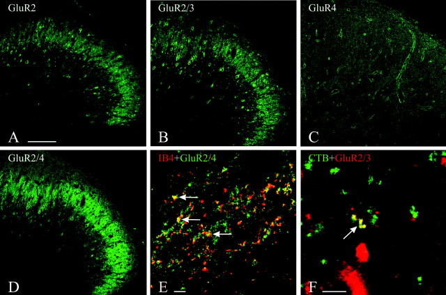
Fig. 4.
A–D, Immunofluorescent staining in rat dorsal horn at L4. The staining for GluR2 (A), GluR2/3 (B), and GluR2/4 (D) is in neuronal perikarya and dense puncta, primarily in lamina II; the staining for GluR4 (C) is punctate and more diffuse. E, F, Double-staining for AMPAR subunits and tracers.E, Staining for GluR2/4 has been combined with IB4 immunofluorescence to detect presynaptic labeling of presumed unmyelinated fiber terminals in lamina II. F, Staining for GluR2/3 has been combined with CTB immunofluorescence to detect presynaptic labeling of presumed myelinated fiber terminals in lamina III. Colocalization is indicated by arrows(E, F). Scale bars, 100 μm.

