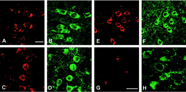
Fig. 11.
Staining of activated MAP kinase family members (in red) and the prolyl isomerase Pin1 (inred) in cerebral cortex from mice of the human P301S tau line. Colocalization with hyperphosphorylated tau protein (ingreen) is shown. A, Anti-activated MAP kinase; C, anti-phospho-JNK; E, anti-phospho-p38; G, anti-Pin1. B, Staining with anti-tau antibody BR134 of the tissue section shown inA. D, F, Staining with anti-tau antibody PHF1 of the tissue sections shown in Cand E. H, Staining with anti-tau antibody AT100 of the tissue section shown in G. Scale bars:A, 15 μm (for A–F);G, 15 μm (for G,H).

