
Fig. 4.
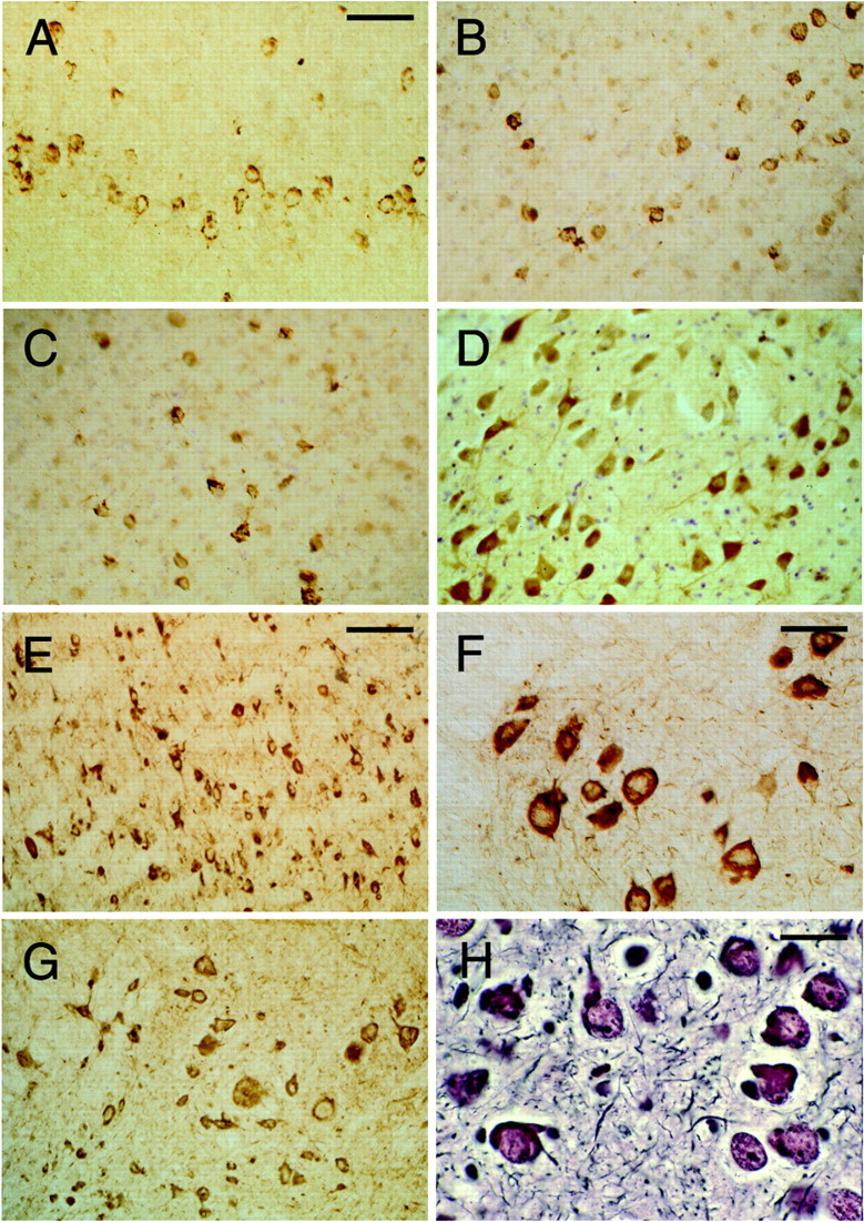
Tau protein immunoreactivity and silver staining in brains and spinal cords from mice of the human P301S tau line. The human-specific, phosphorylation-independent anti-tau antibody T14 was used to stain the cerebral cortex (A) and spinal cord (F). The phosphorylation-dependent anti-tau antibodies AP422 (B) and CP3 (C) were used to stain the cerebral cortex. The phosphorylation-dependent anti-tau antibodies 12E8 (D) and AT180 (E) were used to stain the brainstem. The phosphorylation-dependent anti-tau antibody AT100 was used to stain the spinal cord (G). Bodian silver staining of the amygdala is shown in H. The transgenic mice used were 5 months old. Scale bars: A, 125 μm (forA–D, G); E, 250 μm;F, 100 μm; H, 40 μm.