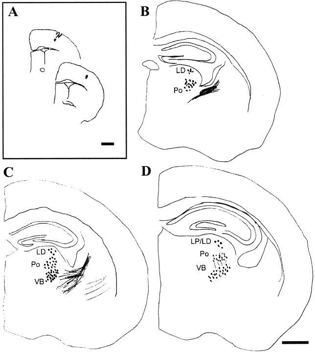
Fig. 3.
Thalamocortical projections in an ephrin-A5-deficient animal. A, Tracer injection site; DiI crystals in the somatosensory cortex were confined to two consecutive 100-μm-thick sections. B–D, Charts of sections with retrogradely labeled cells in the thalamus and thalamocortical axons extending through the internal capsule. Scale bars: A, 1 mm; (in D)B–D, 500 μm.

