
Fig. 2.
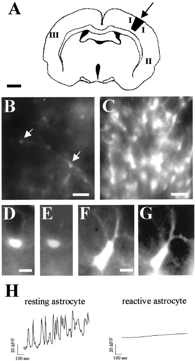
Reactive astrocytes lack spontaneous [Ca2+]i transients. A, Schematic diagram illustrating the location of the stab wound lesion in the parietal cortex (arrow) of P24 GFAP/GFP neocortex and the areas in which spontaneous astrocytic activity was recorded 48 hr after the lesion (I, II, andIII). B, C, Low-magnification photomicrographs showing GFAP/GFP-positive astrocytes (arrows) in areas III (B, resting astrocytes) and I (C, reactive astrocytes). Note that the number of astrocytes and the intensity of GFP fluorescence are markedly increased in astrocytes around the lesion.D–G, Higher-magnification photomicrographs of typical GFAP/GFP-positive resting astrocytes in area III (D, E) and reactive GFAP/GFP-positive astrocytes in area I (F, G). Intense GFP positive-reactive astrocytes have larger somata and hypertrophied processes (F) than resting GFP cells (D). In both resting (E) and reactive (G) astrocytes, the fura-2 indicator was loaded similarly.H, [Ca2+]i profiles over 800 sec of representative GFAP/GFP-positive astrocytes recorded near the lesion site (reactive astrocyte; area I) and in the contralateral hemisphere (resting astrocyte; area III). Scale bars: A, 1 mm; B,C, 40 μm; D–G, 5 μm.