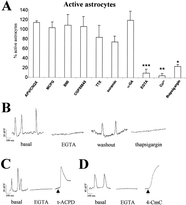
Fig. 3.
Pharmacological analysis of spontaneous [Ca2+]i transients in hippocampal astrocytes. A, Histograms illustrating the percentage of hippocampal P2–P6 GFAP/GFP-positive astrocytes showing spontaneous Ca2+ activity after administration of a range of neurotransmitter receptor antagonists, TTX, and blockers of Ca2+ mobilization. Significant reductions are observed after EGTA, Co2+, and thapsigargin treatments (*p < 0.05; **p < 0.01; ***p < 0.001). Each experimental condition was performed in at least three slices. Data are expressed as percentages of control values (mean ± SEM). B, Representative plots of [Ca2+]ioscillations recorded in the same GFAP/GFP-positive astrocyte from a P6 mouse perfused with normal ACSF (basal and washout), Ca2+-free ACSF (2 mm EGTA, 0 mm [Ca2+]o), and thapsigargin (2 μm). C, D, [Ca2+]i oscillations recorded in P5 GFAP/GFP-positive astrocytes are abolished after perfusion of Ca2+-free ACSF. Addition of t-ACPD and 4-CmC (arrows) to nominally Ca2+-free ACSF caused a transient and a progressive increase in [Ca2+]i, respectively. Abbreviations are defined in Materials and Methods.

