Fig. 4.

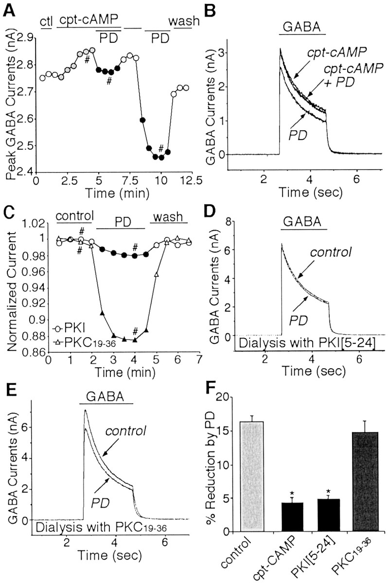
The effect of PD168077 on GABAAcurrents was blocked by PKA activation and occluded by PKA inhibition.A, Plot of peak GABAA currents as a function of time and drug application. In the presence of the membrane-permeable cAMP analog cpt-cAMP (200 μm), PD168077 (PD; 20 μm) failed to reduce GABAA currents. After washing off cpt-cAMP, the effect of PD168077 emerged. B, Representative currenttraces taken from the records used to constructA (at time points denoted by #). C, Plot of peak GABAA currents as a function of time and drug application in neurons dialyzed with PKI[5–24] or PKC19–36. The specific PKA inhibitory peptide PKI[5–24] (20 μm), but not the PKC inhibitory peptide PKC19–36 (20 μm), eliminated PD168077-induced reduction of GABAA currents. D, E, Representative current traces taken from the records used to construct C (at time points denoted by #). F, Cumulative data (means ± SEM) showing the percentage modulation of GABAA currents by PD168077 in the absence (n = 13) or presence of cpt-cAMP (n = 12), PKI[5–24] (n = 8). or PKC19–36 (n = 14). *p < 0.005; ANOVA. ctl, Control.
