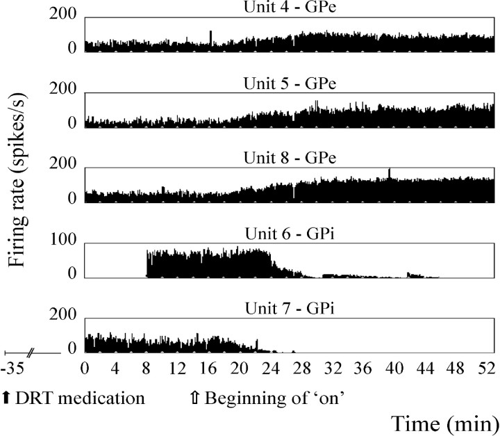
Fig. 2.
Discharge rates of five continuously recorded pallidal neurons during the clinical off–on transition, demonstrating an increase in discharge rate of GPe units and a decrease in GPi units. DRT was administered orally 35 min before onset of recording (solid arrow); clinical off–on transition began after 14 min of recording (open arrow). All units were recorded continuously through minutes 0–52, except for unit 6, which was recorded through minutes 8–46. Thex-axis is time in minutes; y-axis is the firing rate of each unit in spikes/sec.

