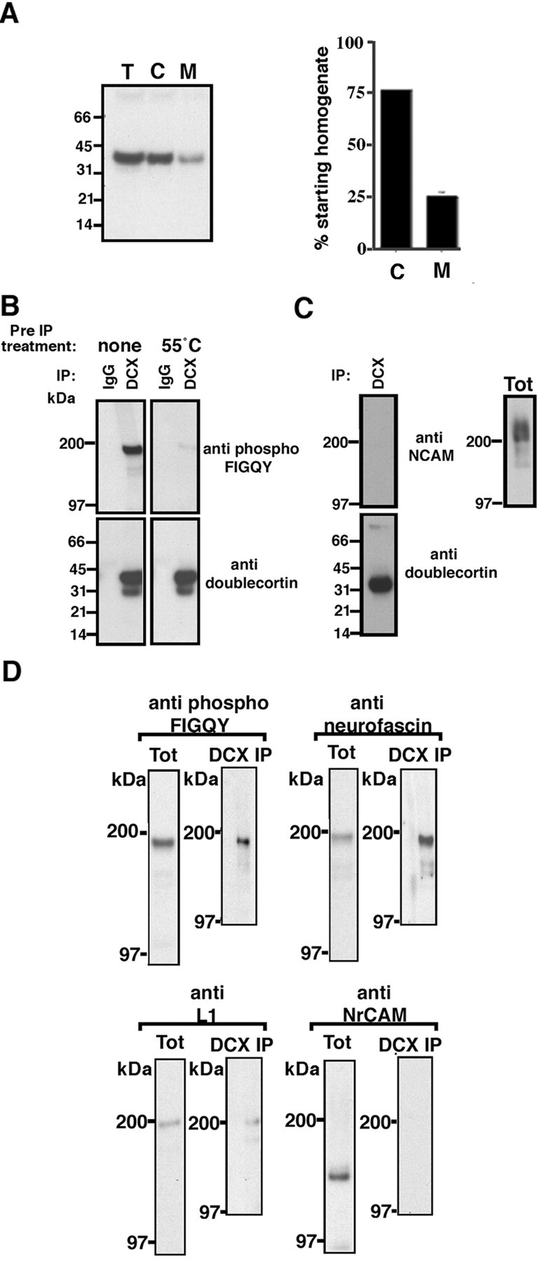
Fig. 2.

Doublecortin and phospho-FIGQY L1 CAMs form a complex in embryonic rat brain.A,Left panel, Immunoblot of equivalent volumes (10 μl) of total embryonic brain homogenate (T), cytosolic fraction (C), and Triton X-100 extract of membrane fraction (M) with a rabbit polyclonal doublecortin antibody. Right panel, Doublecortin in the different fractions (see Materials and Methods) is represented as a percentage of the amount in the starting homogenate (T). B, Immunoblot analysis of immunoprecipitates obtained using doublecortin antibodies from Triton X-100 membrane lysates (Materials and Methods). Antibodies used in immunoprecipitation are shown at thetop of the blots. Antibodies used for immunobloting are shown on the right-hand side of the blots. DCX, Doublecortin; IP, immunoprecipitation. C, Immunoblot analysis of immunoprecipitates obtained using doublecortin antibody as well as total Triton X-100 membrane lysate with anti-NCAM antibody.D, Immunoblot analysis of immunoprecipitates obtained using doublecortin antibody as well as total Triton X-100 membrane lysate with antibodies against neurofascin, L1, and NrCAM. The antibody used for immunoblot is shown on the top of theblots. Tot, Triton X-100 membrane lysate;DCX IP, doublecortin immunoprecipitate.
