Fig. 5.
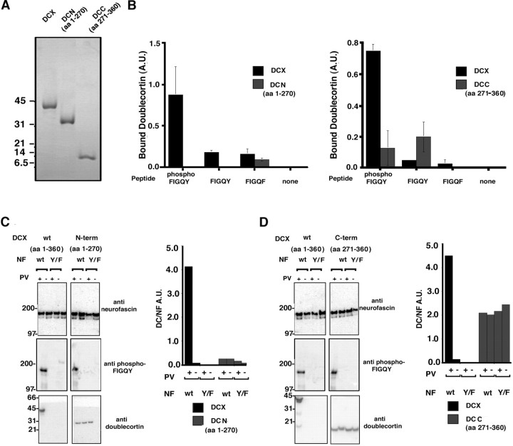
Individual domains of doublecortin fail to bind the phospho-FIGQY motif. A, Coomassie blue-stained gel of purified monodisperse recombinant doublecortin, DCN domain, and DCC domains. B, Peptide binding assay. Thegraph on the left shows the result of assay testing the ability of the DCN domain to bind peptides bearing the phospho-FIGQY motif. Black bars, Doublecortin;gray bars, DCN domain. The graph on theright shows the result of assay testing the ability of the DCC domain to bind peptides bearing the phospho-FIGQY motif.Black bars, Doublecortin; gray bars, DCC domain. Binding is represented as arbitrary units. The peptides used are listed below each bar representing doublecortin binding. C, Binding of native doublecortin and DCN domain to immunoisolated native neurofascin was performed as described (Materials and Methods). The antibodies used for immunoblotting are shown to the right of the blots. The graphical representation of the binding is shown to the right of the blots also. D, Binding of native doublecortin and DCC domain to immunoisolated native neurofascin was performed as described in Materials and Methods. The antibodies used for immunoblotting are shown to the right of theblots. In C and D, the neurofascin used in the binding is also indicated above the blot.wt, Wild type; Y/F, FIGQY/F neurofascin. Pervanadate treatment or lack of it is indicated by + or −, respectively. DCX, Doublecortin; NF, neurofascin; PV, pervanadate. The y-axis represents in arbitrary units the ratio of doublecortin signal to the neurofascin signal after background correction. Black bars, Full-length doublecortin; gray bars, doublecortin domains DCN in C and DCC inD.

