
Fig. 7.
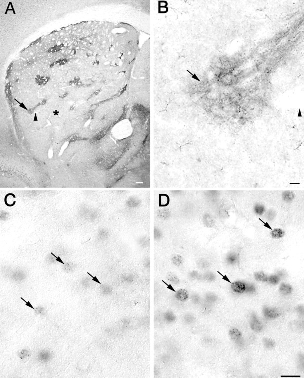
A, Low-power photomicrograph of immunostaining with an MOR-1 antibody in the striatum of a 6-month-old knock-in mouse. Areas positive for MOR-1 correspond to the striosomal compartment (arrow), whereas immunonegative areas correspond to the matrix compartment (asterisk). The arrowhead points to the same blood vessel indicated by the arrowhead in B. Scale bar, 100 μm. B, High-power photomicrograph of MOR-1 immunostaining showing in detail one of the striosomes (arrow) also seen in A atarrow. Scale bar, 10 μm. C,D, High-power photomicrographs of immunostaining with the EM48 antibody in a serially adjacent section to that shown inA and B. C, Striatal neurons with few nuclear microaggregates were found in the matrix compartment (area indicated by the asterisk in A).D, Striatal neurons with numerous microaggregates were located in the striosomal compartment shown in A andB at arrow. Note that only the nucleus of striatal neurons is immunostained. Scale bar (shown in Dfor C and D): 10 μm.