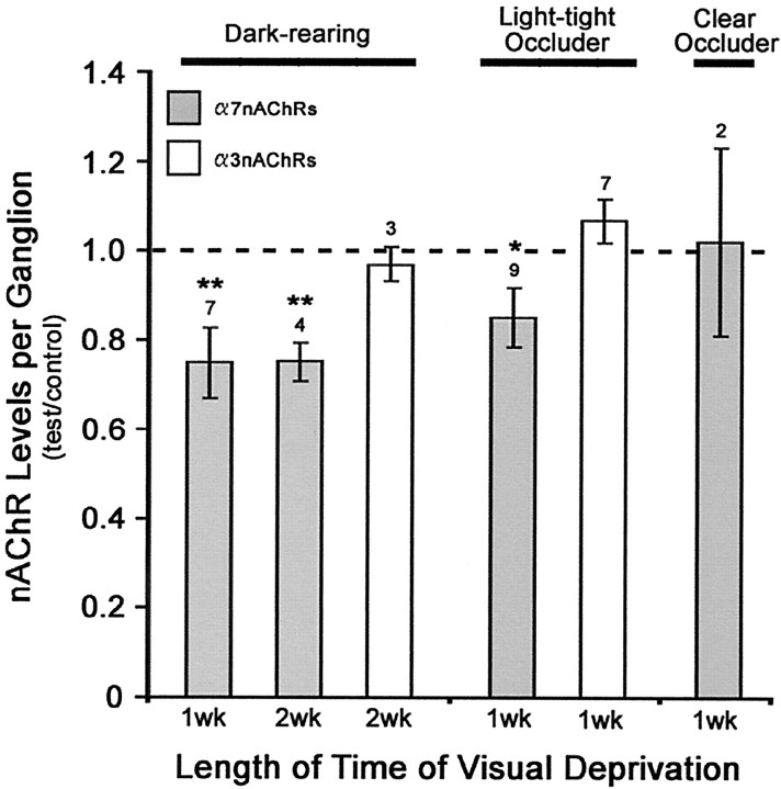
Fig. 4.
Visual deprivation downregulates α7-nAChR levels, but not α3-nAChRs, in newly hatched chick CG neurons. Visual deprivation was used to reduce activity in retinal inputs to CG neurons. Chicks were either dark reared immediately after hatching, or a light-tight occluder was applied over one eye, with the contralateral uncovered eye serving as an internal control. Dark-reared chick CGs are compared with diurnally reared chick CGs at matched ages. To control for nonspecific effects of covering the eye with a plastic goggle, a clear occluder was applied on separate chicks. The total number of α7-nAChRs per ganglion was determined by the specific binding of125I-α-Bgt in ganglionic detergent extracts. Results represent the mean ± SEM of the specific activity (the number of binding sites per ganglionic protein) of the test, with both the mean and SEM values normalized to the mean of the appropriate control. The number of separate determinations is indicated above eachbar. The hatched line represents the control diurnally reared chick CG value. For comparison with α7-nAChRs, the total number of α3-nAChRs was assayed in separate ganglia using 125I-mAb-35. α7-nAChR levels are reduced in visually deprived chick CGs, whereas α3-nAChR levels are not.Asterisks indicate statistically significant differences in the raw data of Bgt-specific activities in test versus control CGs based on the Student's t test for dark-reared chicks and the Student's paired t test for monocular occluded chicks; **p < 0.05 and *p < 0.025, respectively.

