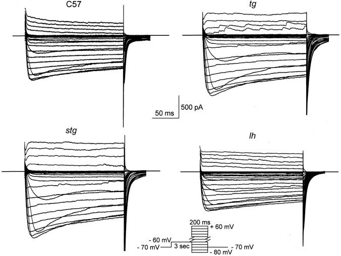
Fig. 4.
Representative superimposed HVA Ca2+ current traces from thalamocortical cells in control, tg, lh, and stgmice. The I–V protocol consisted of a 3 sec prepulse potential at −60 mV, followed by voltage steps (200 msec) ranging from −80 to +60 mV in 5 mV increments, with the holding potential maintained at −70 mV.

