Fig. 6.

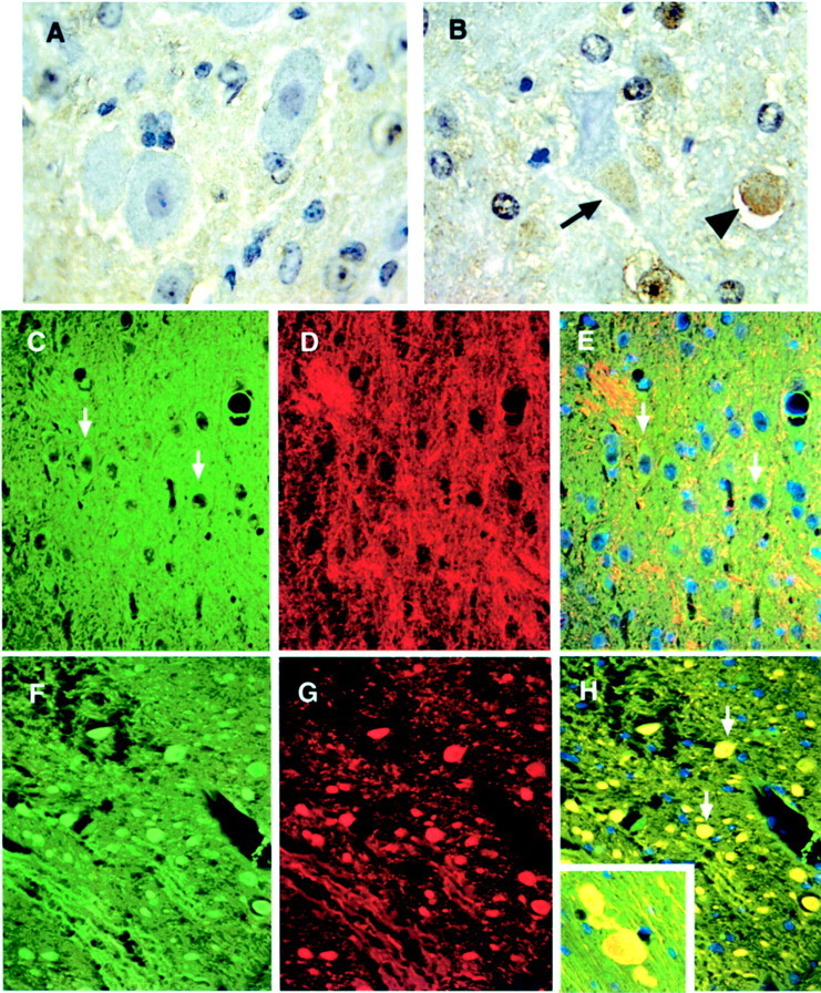
p35 accumulates and colocalizes with phosphorylated NF in axonal spheroids of npc-1 −/− mice. Perfusion-fixed, paraffin-embedded sections from 7-week-old +/+ (A, C–E) and −/− (B, F–H) mice were immunohistochemically labeled with cdk5 (brown) and counterstained with hematoxylin (blue-purple) or immunofluorescently labeled with p35 (Alexa 488, green) and SMI 31 (Cy-3, red) and counterstained with DAPI (blue). cdk5 barely stained neurons in +/+ mice (A, pontine nucleus). In contrast, cdk5 immunoreactivity was increased in the soma of some deformed neurons (arrow) and in the axonal spheroids (arrowhead) in the brainstem of −/− mice (B, pontine nucleus). p35 faintly labeled the soma of neurons in the brainstem of +/+ mice (arrows inC and E, pons) and displayed little colocalization with SMI 31 immunoreactivity (D andmerged image in E). In the −/− mouse brain, intense p35 immunoreactivity was seen in the axonal spheroids (F, pons), which perfectly matched the accumulation of SMI 31 immunoreactivity (G and arrows inmerged image in H).Inset in H, Gigantically enlarged axons containing p35 (green) and SMI 31 (red) immunoreactivity in the cerebral cortex. Magnification: A, B, 100×; C–H, 40×.
