
Fig. 4.
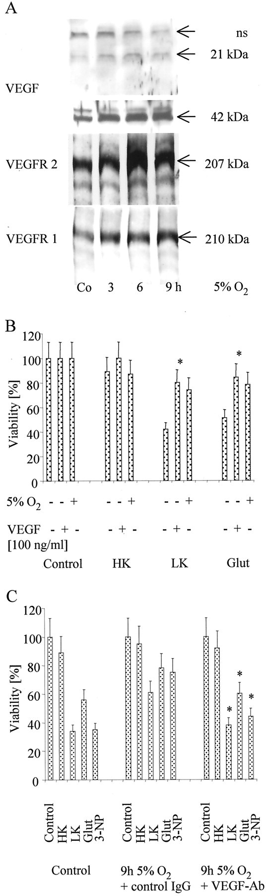
VEGF in hypoxia-induced neuroprotection.A, CGN were cultured at 5% O2 for the indicated intervals. VEGF (monomer at 21 kDa, dimer at 42 kDa; nonspecific bands, ns), VEGFR-2, or VEGFR-1 were assessed by immunoblot. B, CGN were exposed to 5% O2 for 9 hr, reoxygenated for subsequent 24 hr, or pretreated with recombinant VEGF (100 ng/ml) for 24 hr or left untreated, and, subsequently, they were challenged as in Figure2A. Viability was assessed by FDA staining. Data are expressed as means ± SEM from three independent experiments performed in triplicate (*p < 0.05, for the effect of VEGF; ANOVA, followed by Tukey's post hoc test).C, CGN were exposed to 5% O2 as detailed inA, challenged with potassium deprivation, glutamate, or 3-NP as displayed in Figure 2A, and were cotreated with an anti-human IgG control-antibody (10 μg/ml), or with a human neutralizing VEGF-antibody (10 μg/ml) (VEGF-Ab) as indicated. Viability was assessed by FDA staining. Data are expressed as means ± SEM from three independent experiments performed in triplicate (*p< 0.05, for the effect of the VEGF-antibody compared with the control-antibody; ANOVA, followed by Tukey's post hoctest). HK, High K+; LK, low K+; Glut, glutamate.