
Fig. 4.
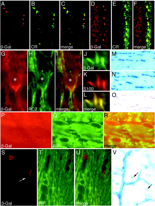
Cajal-Retzius cells, astrocytes, and oligodendrocytes. A–F, Retrosplenial cortex (A–C) and visual cortex (D–F) of P4 Emx1IREScre;R26R mice probed with anti-calretinin and anti-β-Gal.G–I, E12.5 EmxIREScre;R26R cortical plate probed with anti-RC2 and anti-β-Gal.Asterisk indicates nucleus.J–L, Cultured Emx1IREScre;R26R astrocytes probed with anti-β-Gal and anti-S-100.M–O, X-gal-stained adult Emx1IREScre;R26R corpus callosum (M), fimbria (N), and anterior commissure (O). P–U, Emx1IREScre;R26R (P–R) and Emx1IREScre;Z/AP (S–U) corpus callosum probed with anti-β-Gal and anti-Rip. S, Arrow indicates blood vessel. V, Adult Emx1IREScre;Z/AP neocortex. Blood vessels and scattered cells (arrows) are X-gal stained.