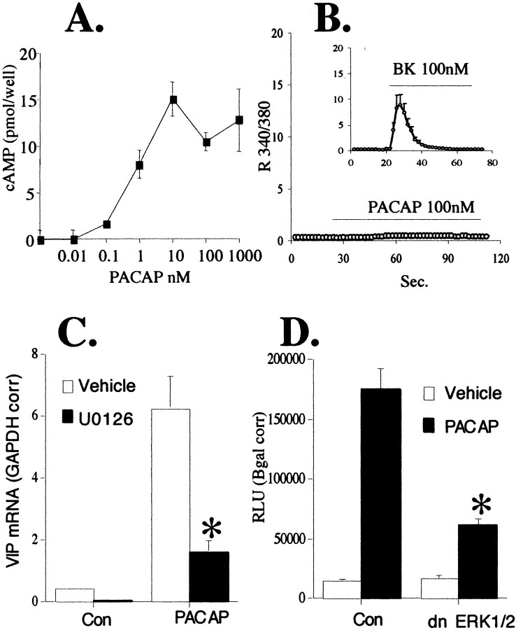
Fig. 9.
PACAP stimulates VIP mRNA elevation in NBFL cells via a MAPK pathway. A, PACAP increases cAMP levels in NBFL cells. NBFL cells were plated in 12-well dishes at 4 × 10⁵ cells per well and allowed to grow to 70% confluency. Cells were harvested 10 min after exposure to increasing concentrations of PACAP-27 from 0.01–1000 nm, and cAMP was measured as described in Materials and Methods. B, PACAP does not increase cytosolic calcium in NBFL cells, whereas bradykinin does. PACAP 100 nm or bradykinin (BK) 100 nm were added to the perfusion chamber over glass coverslipped NBFL cells as described in Materials and Methods. Values shown represent the 340/380 nm ratios. C, PACAP stimulation of VIP mRNA in NBFL cells through a MAPK pathway. NBFL cells grown in six-well plates were harvested for VIP mRNA analysis 6 hr after the addition of 100 nm PACAP-27 with or without 30 μm U0126. Q RT-PCR was performed as described in Materials and Methods. Analysis was performed on 0.2 μg of total RNA and corrected for differences in initial RNA input by dividing by the GAPDH concentrations in the same samples. *Different from vehicle-treated PACAP; p < 0.05. D, dn ERKs inhibit PACAP stimulation of VIP mRNA. NBFL cells were transiently transfected in 12-well plates as described in Materials and Methods. Six hours after the addition of 100 nm PACAP, cells were harvested for luciferase activity. Values are corrected for transfection efficiency by normalizing for β-galactosidase gene expression. *Different from control transfected PACAP-treated cells;p < 0.05.

