
Fig. 6.
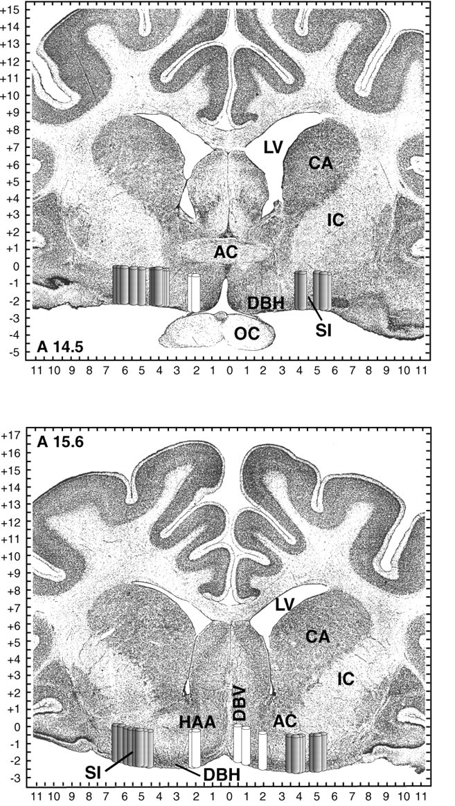
Schematic localization of basal forebrain dialysis sites. Dialysis sites are represented as 2-mm-long, 0.5-mm-widecylinders to indicate the length and diameter of the dialysis probe membrane. Gray cylinders localize 31 lateral basal forebrain dialysis sites in six cats; white cylinders localize seven medial basal forebrain dialysis sites in four cats. Cylinders are shown on coronal atlas plates at 14.5 and 15.6 mm anterior to stereotaxic zero [modified fromBerman and Jones (1982)]. Tick marks on axes indicate 0.5 mm increments. AC, Anterior commissure;CA, caudate; IC, internal capsule;LV, lateral ventricle; OC, optic chiasm.