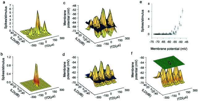
Fig. 2.
Transformations in auditory spatial receptive fields. Receptive fields measured by spiking rate showed either a single tall peak (b) or one tall (main) peak plus smaller side peaks, as in most neurons (a). However, the receptive fields (brown surfaces) measured in subthreshold PSPs had multiple peaks of similar amplitude surrounded by areas of hyperpolarization below the resting potential (bluesurfaces) (c, d). This difference is partly because of a mechanism that converts small differences in membrane potentials to large changes in spike rates (e). In addition, thresholding (green surface represents mean spike threshold measured from membrane potential records) greatly contributes to the isolation of the main peak from other peaks (f). Data in each row came from the same neuron.

