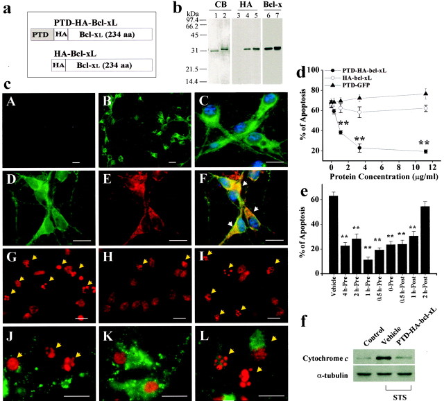
Fig. 1.
The death-suppressing effect of PTD-HA-Bcl-xL in primary cultures of cortical neurons. a, Structures of the Bcl-xL fusion proteins. PTD-HA-Bcl-xL contains both HA tag and the PTD, whereas HA-Bcl-xL does not contain the PTD; the latter served as the control protein in subsequent studies. b, Verification of the Bcl-xL fusion proteins. Coomassie blue (CB) staining shows that the proteins have been purified to near homogeneity. Western blots show that the proteins could be detected using either anti-HA or anti-Bcl-x antibody.c, Representative immunofluorescent images show the transduction and death-suppressing effect in cortical neurons. PTD-HA-Bcl-xL (B, low power; C, high power) but not HA-Bcl-xL (A) transduces into neurons within 15 min of exposure, as determined using HA immunostaining. Immunofluorescence for HA (D) and CCOX IV (E) are partially colocalized (F, overlay), suggesting that a portion of the transduced PTD-HA-Bcl-xL is associated with the mitochondria. The addition of PTD-HA-Bcl-xL (H) but not HA-Bcl-xL (I) reduces the amounts of nuclei showing apoptotic changes (arrows), compared with the vehicle control (G). Immunofluorescence for cytochrome c is preserved in PTD-HA-Bcl-xL-treated neurons (K) but not in HA-Bcl-xL-treated neurons (L) or vehicle-treated neurons (J), consistent with the speculation that PTD-HA-Bcl-xL inhibits STS-induced cytochrome c release.G–L were obtained 24 hr after STS. Scale bar, 20 μm. d, e, PTD-HA-Bcl-xL but not HA-Bcl-xL or PTD-GFP inhibits STS-induced apoptosis in a dose-dependent manner (d). PTD-HA-Bcl-xL was equally effective when it was added to the cultures before or ≤1 hr after STS (e). Apoptosis was quantified after PI staining at 24 hr after STS exposure (0.3 μm). Data are mean ± SE from three independent experiments. **p < 0.01 versus vehicle controls (ANOVA and post hocScheffe's tests). f, Western blots demonstrate that PTD-HA-Bcl-xL treatment attenuated STS-induced cytochromec release to the cytoplasm in cultured neurons. Eachlane contains 20 μg of cytosolic protein, prepared from noninduced (control) or STS-induced (0.3 μm for 6 hr) neurons, respectively. The blots are representative of three independent experiments with similar results.

