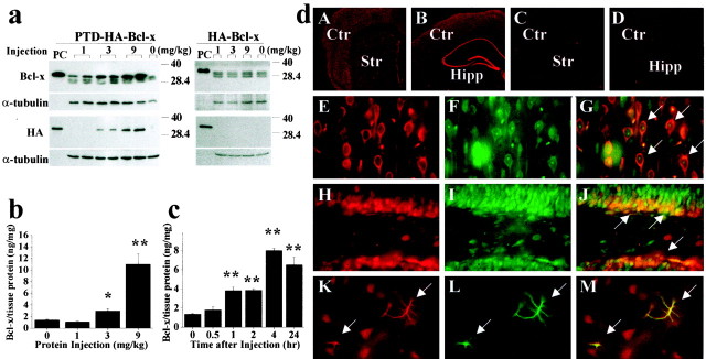
Fig. 2.
In vivo protein transduction of PTD-HA-Bcl-xL in murine brain. a, Western blotting with either the anti-Bcl-x or anti-HA antibody detects the transduction of PTD-HA-Bcl-xL into the cortex 4 hr after intraperitoneal injection of the protein at various doses (left); the HA-Bcl-xL protein lacking PTD failed to transduce across the blood–brain barrier (right). In all blots, the fusion proteins serve as the positive controls (PC). Immunoblotting of α-tubulin serves as a control for sample loadings (30 μg perlane). Note that the endogenous Bcl-x proteins are detectable in all brains. b, c, Quantitation of PTD-HA-Bcl-xL transduction in the brain by ELISA. Intraperitoneal injection of PTD-HA-Bcl-xL results in dose-dependent (b, 4 hr after injection) and time-dependent (c, injection at the dose of 9 mg/kg) protein transduction into the murine cerebral cortex. Data are mean ± SE (three animals per group). *p < 0.05 versus controls; **p < 0.01 versus controls. d, Immunofluorescent detection (using anti-HA antibody) of PTD-HA-Bcl-xL transduction in the cortex/caudate (A) and hippocampal (Hipp) formation (B) 4 hr after intraperitoneal injection; the immunofluorescence is lost after the primary antibody was preabsorbed with the fusion protein (C). Injection of HA-Bcl-xL serves as the negative control (Ctr) (D). In the cerebral parenchyma, most of the cells transduced with PTD-HA-Bcl-xL (HA immunoreactive; E, H, red) are neurons, being immunoreactive for the neuronal markers NSE (F, arrows in G; cortex) or NeuN (I, arrows in J; hippocampal dentate). Some astrocytes (GFAP-immunoreactive;L, green) in the caudate are also transduced (arrows in K–M).Str, Striatum.

