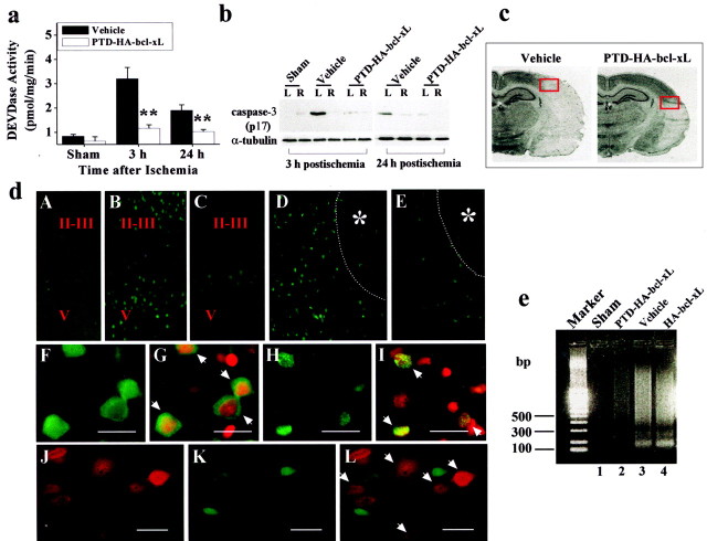
Fig. 4.
PTD-HA-Bcl-xL inhibits an apoptotic component of ischemic neuronal death. a, Effects of PTD-HA-Bcl-xL on caspase-3-like protease activity in the brain at 3 and 24 hr after ischemia, as determined in the cortical cell extracts using the fluorogenic substrate DEVD-AFC. Data are mean ± SE; n = 5 per group. **p < 0.01 versus vehicle treatment. b, Attenuation of caspase-3 activation after ischemia by PTD-HA-Bcl-xL, as demonstrated by Western blotting. The active (p17) caspase-3 fragment is increased in the ischemic hemisphere (L) compared with the nonischemic side (R) in vehicle-treated but not in PTD-HA-Bcl-xL-treated brains. The blots are representative of three independent experiments with similar results. c, Coronal sections obtained from vehicle-treated and PTD-HA-Bcl-xL-treated brains, respectively, 24 hr after ischemia. The red boxes mark the approximate locations from which the immunofluorescent images in c were obtained.d, Representative immunofluorescent images show that PTD-HA-Bcl-xL attenuates caspase-3 activation after ischemia. Immunostaining was accomplished using the antibody recognizing the active caspase-3. A–E, Low power (25×):A, Nonischemic cortex. B, At 3 hr after ischemia, caspase-3-immunoreactive cells are present primarily in layers I-II and V of the cortex. C, The mounts of caspase-3-immunoreactive cells are decreased in PTD-HA-Bcl-xL-treated brain. D, 24 hr after ischemia, caspase-3-immunoreactive cells are localized in the border zone of the infarction (*). Thedotted lines in D and E mark the estimated infarct border. E, Few caspase-3-immunoreactive cells are present in PTD-HA-Bcl-xL-treated brain. F–I, high power (400×). Scale bar, 20 μm. F, At 3 hr after ischemia (vehicle treated), caspase-3 immunoreactivity primarily shows a cytosolic location. G, As indicated by the arrows, the cells in F counterstained with the DNA dye PI.H, At 24 hr after ischemia, caspase-3 immunoreactivity is localized in the nucleus. I, As indicated by thearrows, the cells in H counterstained with PI. J–L, Double-label immunofluorescence of PTD-HA-Bcl-xL (J) and caspase-3 (K) in the border zone of the cortical infarction at 24 hr after ischemia (PTD-HA-Bcl-xL-treated brain); Lis the overlay of J and K. Note that there is no colocalization of caspase-3 and PTD-HA-Bcl-xL in neurons (L, arrows indicate protein-transduced neurons). e, Apoptotic DNA fragmentation is attenuated by PTD-HA-Bcl-xL after ischemia, as demonstrated by DNA electrophoresis. The DNA was prepared from the cortical tissues 72 hr after ischemia, using the brains pretreated with vehicle, PTD-HA-Bcl-xL, or HA-Bcl-xL. The gel is representative of two independent experiments with similar results.

