Fig. 5.
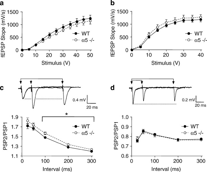
Synaptic strength in hippocampal brain slices of α5 −/− mice compared with its effect on paired-pulse facilitation in CA1 region and paired-pulse depression in the dentate gyrus. In all figures the filled circles represent data from age-matched WT mice, and open circles are from α5 −/− mice. No significant difference was found in the maximal fEPSP amplitude in the CA1 region. a, Forty-six slices from 16 WT controls and 48 slices from 16 α5 −/− mice or dentate gyrus.b, Forty-one slices from 16 WT controls and 42 slices from 16 α5 −/− mice. In the CA1 region the amplitude of fEPSPs was significantly enhanced during paired-pulse stimulus intervals of 100–300 msec. c, Filled circles, 46 slices from 16 WT controls; open circles, 48 slices from 16 α5 −/− mice. In comparison, paired-pulse depression of fEPSPs in the dentate gyrus remained unaffected. d, Filled circles, 41 slices from 16 WT controls; open circles, 42 slices from 16 α5 −/− mice.

