
Fig. 4.
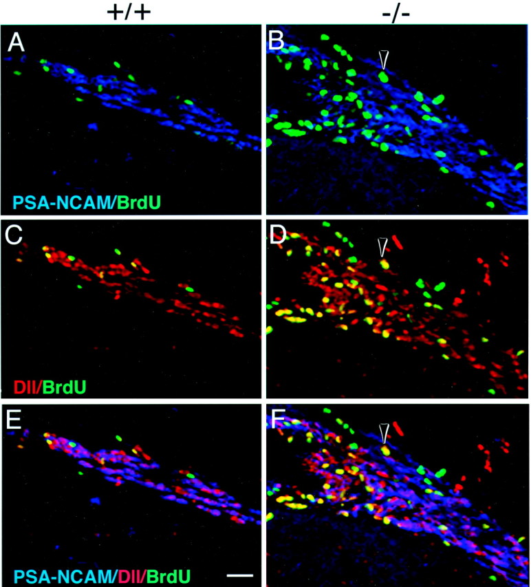
Identification of proliferating type C cells in p27Kip1-null mice. Triple immunofluorescence of frozen coronal sections obtained from the SVZ of wild-type (+/+, A, C, E) and p27Kip1-null (−/−, B, D, F) mice and stained for BrdU (green nuclei), PSA-NCAM (blue immunofluorescence), and Dll (redimmunofluorescence). The arrowhead indicates a cell that is BrdU+/PSA-NCAM− (B, F) but Dll+ (D, F) and therefore identified as a proliferating type C cell. Note that in the mutant, the accumulation of proliferating C cells close to the ventricular lumen (on theleft of each panel) displaces the PSA-NCAM+ type A cells laterally. Scale bar, 100 μm.