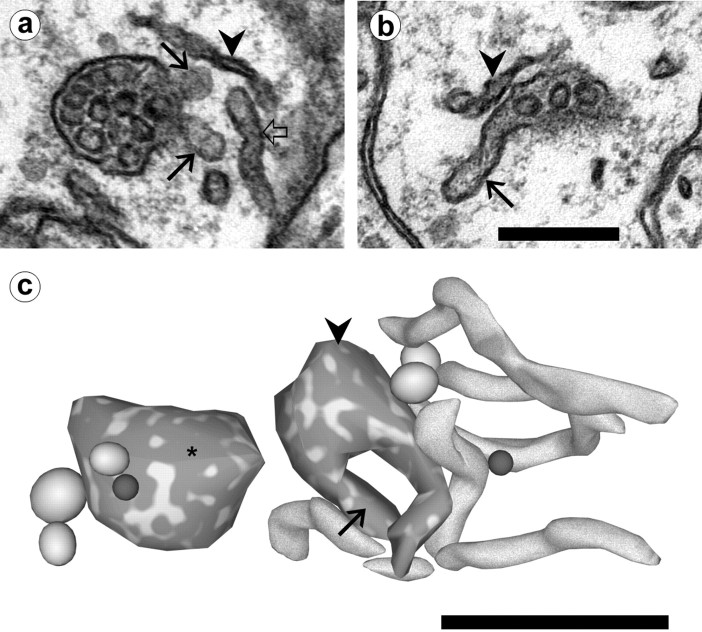
Fig. 5.
Multivesicular body/tubule complexes in dendrites.a, An MVB with attached tubules (arrows). Compare the flattened appearance of SER (arrowhead) with the more cylindrical shape of an unattached tubule (open arrow) located nearby. b, A tubule (arrow) is in continuity with an MVB only partially visible on this section. SER is located nearby (arrowhead). Scale bar (shown in b fora and b): 0.25 μm. c, Reconstruction of the MVB–tubule complex of band associated compartments. The tubule (arrow) visible in the electron micrograph is continuous with a round MVB (arrowhead) that has a second tubular extension. Several additional tubular compartments are located adjacent to this structure. An isolated MVB (∗) nearby does not have any tubular extensions. Also reconstructed are several large, ellipsoidal vesicles (gray) and two spherical small vesicles (dark gray). Scale bar, 0.5 μm.

