
Fig. 1.
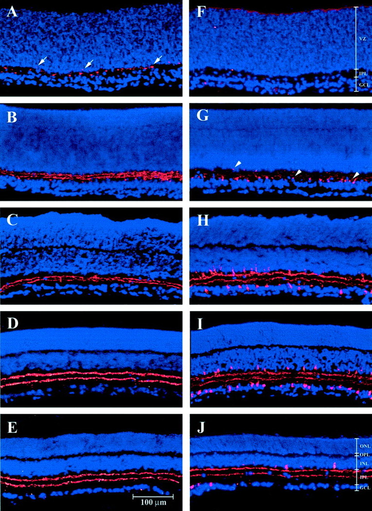
Cholinergic amacrine cells in the developing rat retina. The images are vertical sections of retinas with the inner, or corneal, side at the bottom of the section. VAChT antibody labeling is shown on the left, with ChAT labeling on the right (red). Sections are counterstained with DAPI nuclear stain (blue). Fromtop to bottom, the images show protein expression on the day of birth (P0) (A,F), P2 (B, G), P6 (C, H), P12 (D,I), and in the adult (E,J). The layers of the immature postnatal retina (F) include the ventricular zone (VZ), the inner plexiform layer (IPL), and the ganglion cell layer (GCL). In the mature retina (J), the layers are the outer nuclear, or photoreceptor, layer (ONL), the outer plexiform layer (OPL), the inner nuclear layer (INL), the IPL, and the GCL. Arrows show the earliest VAChT staining at P0 (A), and arrowheadsshow the earliest ChAT staining at P2 (G).