
Fig. 1.
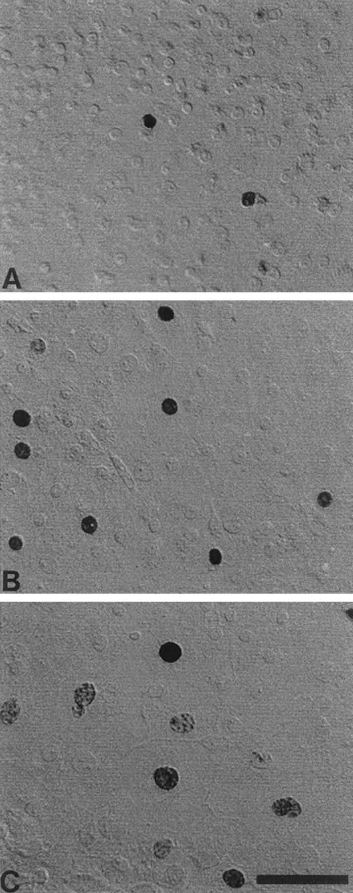
Morphology of supporting cells in selected regions of epithelial cultures. Cultured cells grew as confluent monolayers on fibronectin and laminin substrates. Photographs illustrate the high-density (A), moderate-density (B), and low-density (C) regions of the cultures. Darkly stained BrdU+ nuclei were present in all regions, but the relative numbers of BrdU+ cells were higher in regions of low cell density. Scale bar, 50 μm.