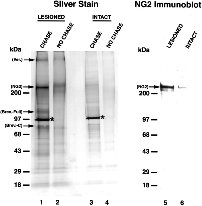
Fig. 1.
Silver stain and immunoblot analysis of total soluble CSPG proteoglycans 2 weeks after SCI. Silver Stain, Lanes 1–4; Immunoblot, lanes 5, 6;lane 1,visualization of prominent core CSPG proteoglycans, 2 weeks after SCI. Core proteoglycans are matched to previously established molecular weights: versican, ∼400 kDa; NG2, ∼300 kDa; brevican full-length, ∼145 kDa; and brevican C-terminal fragment, ∼80 kDa. Use of an 8–16% SDS stacking gel allows for comparison of relative levels of proteoglycan expression, demonstrating that NG2 is a major CSPG species after injury. The asterisk denotes the chondroitinase ABC (CHASE) enzyme added after CSPG isolation.Lane 2, CHASE omission control. A diffuse band corresponding to NG2 runs at a higher level in the gel because of chondroitin sulfates attached to the core protein. CHASE digestion of these sugars allows for clear visualization of the core proteoglycans.Lanes 3, 4, Samples from control tissue. Note the low-level, constitutive expression of a number of CSPGs in lane 3. Lanes 5, 6, NG2 immunoblot shows strong upregulation of NG2 after injury and demonstrates that this proteoglycan runs at the same molecular weight (∼300 kDa) as the band identified with silver stain analysis, confirming the identity of this band as NG2.

