
Fig. 7.
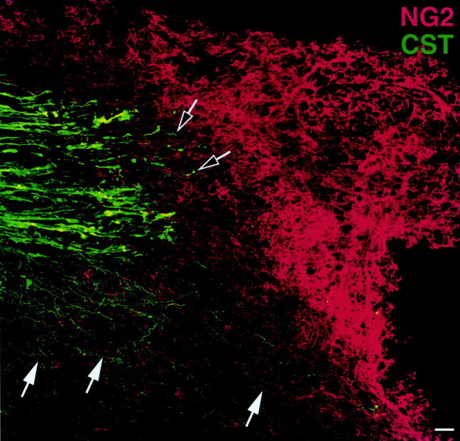
Expression of NG2 in relation to transected corticospinal axons. Confocal analysis of BDA-traced transected CST axons demonstrates deposition of NG2 at the transected ends of the CST axons 7 d after injury. Black and white arrowsindicate that very few CST axons are present in the region of dense NG2 labeling. Notably, sprouting of CST axons (white arrows) occurs in the underlying gray matter where NG2 expression is least prominent. Thus, NG2 deposition could limit axonal regeneration after CNS injury. This figure is a composite of 12 individual images. Scale bar, 39 μm.