
Fig. 4.
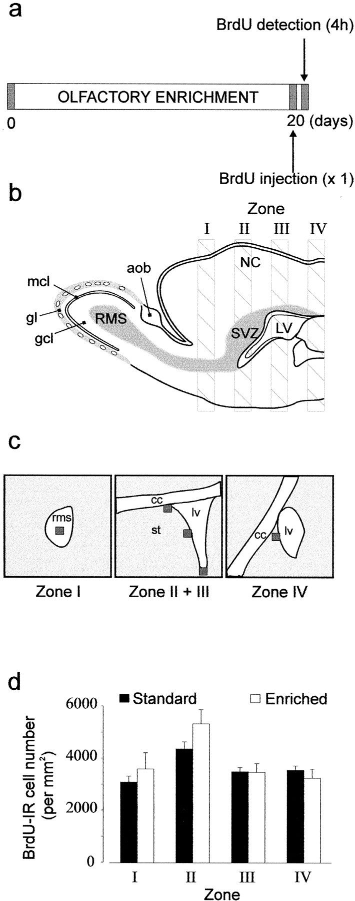
Quantification of proliferation by counting BrdU-labeled cells in enriched (n = 4) and standard (n = 5) mice throughout the rostrocaudal axis extending from the RMS to the caudal part of the lateral ventricle.a, Protocol used to study the proliferation of adult-generated cells labeled with BrdU. A unique intraperitoneal BrdU injection was done 20 d after animals were assigned to their respective groups (day 0), and mice were killed 4 hr after BrdU injection. b, Diagram of a parasagittal view showing the four sampling zones along a rostrocaudal axis. c, Schema representing the RMS, the lateral ventricle (lv), the corpus callosum (cc), and the striatum (st). Squares (50 × 50 μm) indicate three analyzed areas (ventral, lateral, and dorsal) for BrdU-IR cells in Zone II + III of the lateral ventricle wall and one analyzed area both in the RMS and in Zone IV. d, Mean number of BrdU-IR cells per millimeters squared in each defined zone for standard and enriched mice.