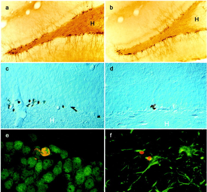
Fig. 2.

Illustration of PSA-NCAM- and BrdU-labeled cells in the dentate gyrus. Microphotography of PSA-NCAM staining in a control animal (a) and in an animal self-administering 0.04 mg/kg per infusion of nicotine (b). Microphotography of BrdU staining in a control animal (c) and in an animal self-administering 0.08 mg/kg per infusion of nicotine (d). Optical section (0.7 μm) obtained by confocal microscopy showing that BrdU-stained cells (red nuclear stain, CY3) were double-stained with the neuronal marker NeuN (green stain, Alexa 488) (e). In contrast, very few BrdU-stained cells (red nuclear stain) also expressed the astroglial marker GFAP (green stain, f). H,Hilus.
