
Fig. 4.
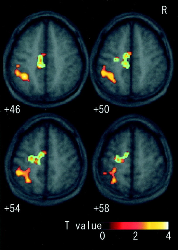
Fields activated by motor imagery in sections fromz +46 to +58 superimposed on the mean MRI of all subjects. When imagery was contrasted with rest, clusters in the contralateral (left) CMA, SMA, PMD, and intraparietal sulcus area extending into postcentral sulcus were significantly activated. Blue areas were the identical sections that were also significantly activated as the common field [(imagery + illusion) vs (rest + vibration)]. CMA, Cingulate motor area; SMA, supplementary motor area; PMD, dorsal premotor cortex.