
Fig. 5.
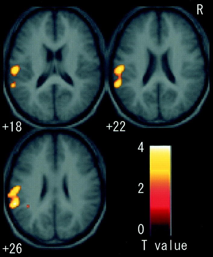
Fields activated by motor imagery in sections fromz +18 to +26. When imagery was contrasted with rest, the cluster in the left parietal operculum was significantly activated. There was no common active field active in this region.
Official websites use .gov
A
.gov website belongs to an official
government organization in the United States.
Secure .gov websites use HTTPS
A lock (
) or https:// means you've safely
connected to the .gov website. Share sensitive
information only on official, secure websites.
Fields activated by motor imagery in sections fromz +18 to +26. When imagery was contrasted with rest, the cluster in the left parietal operculum was significantly activated. There was no common active field active in this region.