
Fig. 1.
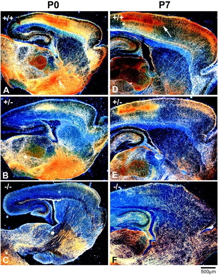
5-HT axons fail to innervate the cortex and hippocampus in neonatal GAP43−/− mice. Low-magnification dark-field photomicrographs of parasagittal sections from P0 (A–C) and P7 (D–F) mice show bright serotonin-immunoreactive axons on a dark background. At P0, 5-HT axons in WT mice are present throughout the brain and densely innervate the thalamus, basal forebrain, and frontal and parietal areas of the cortex (A). The density of 5-HT axons in GAP43−/− mice is dramatically reduced in most brain regions compared with WT mice at P0 (C). The density of 5-HT axons increases in the thalamus, cortex, and hippocampus of WT mice over the next week. At P7, distinct patches of serotonin immunoreactivity are visible in layer IV of the parietal cortex (arrow,D). In GAP43−/− mice at P7, the density of 5-HT axons in subcortical regions is similar to that in WT mice, but very few 5-HT axons are observed in the cortex and hippocampus (F). In GAP43+/− mice at P0 (B) and P7 (E), 5-HT axon density in the hippocampus and cortex is intermediate between that in GAP43−/− and WT mice. Scale bar, 500 μm.