
Fig. 3.
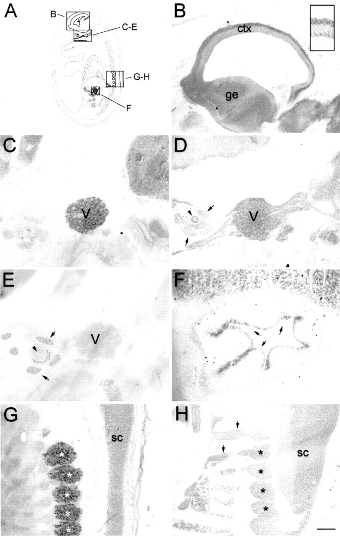
Nogo expression in the E16 rat embryo.In situ hybridization with the nogo-Aprobe revealed strong expression in the mantle layer of postmitotic neurons in the developing forebrain cortex (B,inset). In the trigeminal ganglion (C, D;V), high levels of Nogo-A were detected (C, nogo-A probe; D, AS 472). Nogo-A protein was also found in the trigeminal nerve fibers, extraocular muscles (arrows), and optic nerve (arrowhead). Note that Nogo-C (AS 818; E) was only present in extraocular muscles (arrows) but not in the trigeminal ganglion or optic nerve. Strong expression of Nogo-C was found with AS 818 in intestinal epithelium (F,arrows). G, H, Nogo-A mRNA (G) and protein (H) expression in the spinal cord (sc) and DRG (asterisks). Again, protein expression revealed by AS 472 was also found in nerve fibers (H, arrows), whereas mRNA expression (nogo-A probe) was restricted to neuronal cell bodies in the DRG and spinal cord. The level of mRNA expression in DRG was higher than in the spinal cord; however, protein levels were comparable. Scale bar: B–E, G, H, 550 μm;F, B, inset, 140 μm.