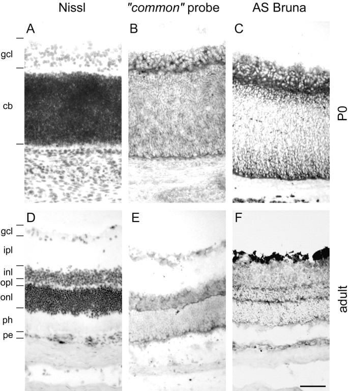
Fig. 8.

Nogo expression in the retina. At birth (A–C), strong expression of Nogo-A/B mRNA (B, common probe) and protein (C, AS Bruna) in the ganglion cell layer (gcl) as well as the cytoblast layer (cb) was observed. Expression was highest at the interface between the ganglion cell layer and cytoblast layer. The ganglion cells were also Nogo-A/B-positive in the adult (D–F), and both inner and outer nuclear layers (inl, onl) were expressing mRNA (E, common probe) and protein (F, AS Bruna). Nogo-A/B protein seemed to be targeted to neurites, because the inner and outer plexiform layers (ipl, opl) were positive for AS Bruna (F). A, D, Nissl staining. Scale bar, 70 μm.
